Some of the most important moments in cancer research happen in seconds. A new imaging technology developed by MUSC startup OncoBLAZE lets researchers see those moments as they happen.
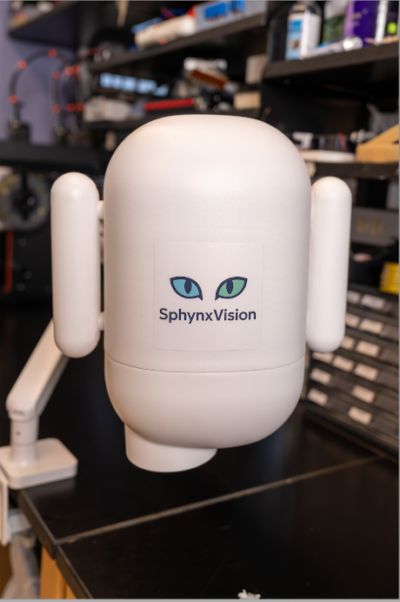
Called SphynxVision, the imaging system is designed for early-stage research conducted before being tested in patients. It allows scientists to capture high-resolution fluorescence images in real time under normal room lighting. That sets it apart from conventional systems, which require samples to be placed inside closed, dark chambers, limiting researchers’ ability to see what is happening during an experiment or procedure.
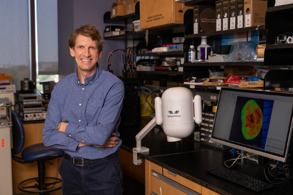
SphynxVision removes that barrier with an open design that allows an experiment to be observed as it is performed. That means researchers could watch where a drug travels as it is injected, track what is happening during active surgery or observe how tissue responds to other interventions—potentially revealing changes they might otherwise miss.
“SphynxVision was built around the experiment, not around the imaging chamber,” said MUSC Hollings Cancer Center researcher Dieter Haemmerich, PhD, CEO of OncoBLAZE. “Researchers often need to see what is happening while they perform surgeries or treat subjects. Our system makes that possible in a flexible, bench-side system.”
OncoBLAZE unveiled SphynxVision at the World Molecular Imaging Congress in Salt Lake City in September, marking an important step toward bringing the company’s first commercial product to market. There, the team recruited a small group of laboratories to their Pioneer Program to test an early version of the system.
Seeing biology as it happens
Fluorescence imaging gives researchers a way to make specific cells, molecules, or drugs “light up.” By attaching fluorescent labels to what they want to track, researchers can see, for example, where cancer cells are located or where a drug travels in a living being.
But there is a catch: Ordinary room light can drown out that fluorescent signal.
Conventional systems solve that problem by putting the subject inside a dark chamber. While that works well for many experiments, it limits what researchers can do while imaging. They may have to take an image, stop while a procedure is performed and then return to the chamber to take another image. That provides snapshots of what happened before and after—but leaves out what happened in between.
SphynxVision was built around the experiment, not around the imaging chamber. Researchers often need to see what is happening while they perform surgeries or treat subjects. Our system makes that possible in a flexible, bench-side system.
Dieter Haemmerich
SphynxVision takes a different approach. Its light source is about 100 times brighter than those used in conventional systems, creating a much stronger fluorescent signal. Specialized image processing then filters out remaining interference from room light. Together, these proprietary technologies allow researchers to see fluorescence more clearly without putting their experiments in the dark.
The system’s speed and flexibility give researchers a more complete picture. SphynxVision can capture multiple images per second, tracking biological changes in real time while greatly reducing the blurring that movement can cause. Its higher resolution can also reveal remarkably small details, such as tiny blood vessels or clusters of cancer cells, that may be difficult or impossible to see with conventional systems.
Seeing an experiment as it happens gives researchers information that a final image alone cannot provide.
Consider a cancer drug. Researchers use fluorescence imaging to track where the drug ends up. With SphynxVision, they can go even further by watching the drug move through the body as it is delivered, revealing where it goes, when it arrives and how its distribution changes over time.
That real-time view is valuable when changes happen within seconds. It could also help researchers develop new technologies for cancer surgery. Haemmerich pointed to work with Hollings head and neck cancer surgeon Dauren Adilbay, MD, PhD, who is using SphynxVision in preclinical studies to develop and test a new fluorescent contrast agent.
An unexpected innovation
Haemmerich and colleagues did not set out to reinvent preclinical imaging.
SphynxVision grew from more than a decade of research at MUSC and Hollings focused on delivering chemotherapy more precisely to tumors while limiting how much reaches other parts of the body. To make that approach work, they needed to see where the chemotherapy was going as it was delivered.
“There wasn’t a system able to do that,” Haemmerich said. “That’s why we developed this.”
What began as a solution to a problem in their own lab soon revealed much broader potential. The team realized the same fluorescence imaging technology could give other researchers a new way to observe experiments as they unfold.
That realization became SphynxVision. Over the past three years, the team has worked to turn its laboratory prototype into a reliable, accessible system that other scientists can use.
Bringing that technology out of the laboratory is where OncoBLAZE comes in. Co-founded by Haemmerich and Christian Rossmann, PhD, OncoBLAZE commercializes technology developed at MUSC and is based in MUSC’s Blue Sky Labs incubator, an innovation hub that supports early-stage life science companies. Federal funding from the National Institutes of Health, National Science Foundation, and South Carolina Research Authority also supported the technology’s development.
OncoBLAZE has already earned statewide recognition for its work. The startup was one of three finalists for the Technology Development Award at the 2025 InnoVision Awards, which recognize innovation in science, technology, and business across South Carolina.
Taking SphynxVision beyond the lab
The next step is putting the technology into the hands of other researchers.
We hope SphynxVision allows researchers to see things they haven’t been able to see before—to image experiments as they’re happening and ultimately answer questions that current imaging systems can’t.
Dieter Haemmerich
At September’s World Molecular Imaging Congress, OncoBLAZE recruited researchers for its SphynxVision Pioneer Program. The company will place early versions of the system in four or five laboratories, where scientists will test them with real-world research and share feedback ahead of a broader commercial launch.
For Haemmerich, success will ultimately be measured by more than how well the technology performs. It will be about the discoveries it makes possible.
“We hope SphynxVision allows researchers to see things they haven’t been able to see before—to image experiments as they’re happening and ultimately answer questions that current imaging systems can’t.”